

Interactive CT & MRI Anat.Lite

Descripción de Interactive CT & MRI Anat.Lite
★Lite version★
This is the free Lite version of "Interactive CT and MRI Anatomy".
The function is restricted.
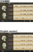
You can only see the transverse CT images of the head.
Please check the operation before purchasing the full version.
★ Details ★
This application is developed for medical students, interns, residents, doctors, nurses, and radiology technicians to understand the essential anatomical terms of the body.
You can learn anatomy by answering the terms by step-to-step questions using a total of 241 CT and MRI images.
A total of 17 images of 3D-CT, MRA and plain X-ray film(particularly the extremities) are included as references.
Other reference images include coronary artery segments defined by the American Heart Association(AHA), pulmonary segments, and liver segments(according to Couinaud classification).
You can enlarge all the images by simple manipulation.
★ Major functions ★
There are 4 major functions.
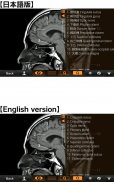
-1) Anatomical mode
Anatomical terms are overlaid on the images.
It can be used as the anatomical atlas.
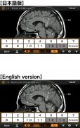
-2) Quiz mode type 1
You select the part of the image by using anatomical term.
Questions will basically appear randomly.
-3) Quiz mode type 2
You select the anatomical term by the part of the image.
Questions will basically appear randomly.
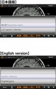
-4) Index
You can find the specific images by using anatomical terms.
★ Intended users ★
-Medical students
-Interns and residents
-Doctrors
-Nurses
-Radiology technicians
-All those who are intrested in CT and MRI anatomy
★ Images(a total of 258 images) ★
Images basically include horizontal, coronal, and sagital planes.
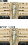
-Head(36 images including CTA and 3D-CT)
-Neck(24 images)
-Spine(19 images including plain X-ray films)
-Chest(61 images including 3D-CT images)
-Abdomen (37 images)
-Pelves: male (9 images)
-Pelvis: female (11 images)
-Extremities (shoulder, hand, elbow, hip joint, knee, foot) (61 images including plain X-ray films)
Editors
Toshiaki Nitori, M.D. (Professor of Radiology, Kyorin University, School of Medicine)
Yasuo Sasaki, M.D. (Manager of diagnostic radiology, Iwate Prefectural Central Hospital)
</div> <div jsname="WJz9Hc" style="display:none">★ ★ versión Lite
Esta es la versión gratuita Lite de "Interactivo TC y la RM Anatomy".
La función está restringida.
Sólo puede ver las imágenes de la TC transversales de la cabeza.
Por favor verifique la operación antes de comprar la versión completa.
★ ★ Detalles
Esta aplicación está desarrollada para estudiantes, internos, residentes, médicos, enfermeras y técnicos de radiología médica para entender los términos anatómicos esenciales del cuerpo.
Usted puede aprender anatomía, respondiendo a los términos de preguntas de paso a paso con un total de 241 imágenes de TC y RM.
Un total de 17 imágenes de 3D-CT, MRA y radiografía simple de rayos X (en particular las extremidades) se incluyen como referencia.
Otras imágenes de referencia incluyen segmentos coronarios arteriales definidos por la American Heart Association (AHA), segmentos pulmonares, y segmentos de hígado (según la clasificación Couinaud).
Usted puede ampliar todas las imágenes mediante la manipulación sencilla.
★ ★ funciones principales
Hay 4 funciones principales.
-1) Modo anatómico
Términos anatómicos se superponen sobre las imágenes.
Se puede utilizar como los atlas anatómicos.
-2) Modo Prueba tipo 1
Seleccione la parte de la imagen mediante el uso de término anatómico.
Las preguntas serán básicamente aparecerá aleatoriamente.
-3) Modo Prueba tipo 2
Usted selecciona el término anatómico por la parte de la imagen.
Las preguntas serán básicamente aparecerá aleatoriamente.
-4) Índice
Puede encontrar las imágenes específicas mediante el uso de términos anatómicos.
★ ★ usuarios previstos
Estudiantes -Médico
-Interns Y residentes
-Doctrors
-Enfermeras
Técnicos -Radiology
-Todos Aquellos que se interese TC y la RM la anatomía
★ Imágenes (un total de 258 imágenes) ★
Imágenes básicamente incluyen planos horizontales, coronal, sagital y.
-Jefe (36 imágenes incluyendo CTA y 3D-CT)
-Neck (24 imágenes)
-Spine (19 imágenes incluyendo películas de rayos X de civil)
-Chest (61 imágenes incluyendo imágenes 3D-CT)
-Abdomen (37 imágenes)
-Pelves: Masculino (9 imágenes)
-Pelvis: Femenino (11 imágenes)
-Extremities (Hombro, mano, codo, cadera, rodilla, pie) (61 imágenes incluyendo películas de rayos X de civil)
Editores
Toshiaki Nitori, MD (Profesor de Radiología de la Universidad Kyorin, Facultad de Medicina)
Yasuo Sasaki, MD (Gerente de la radiología diagnóstica, Iwate Hospital Central de la Prefectura)</div> <div class="show-more-end">


